Urgent Message: The azygos lobe may be apparent on a chest x-ray and is a relatively uncommon, yet benign finding, that is important to distinguish from other etiologies. The azygos lobe is an embryological remnant and is usually incidental in nature.
Citation: Richmond C, Mancuso A. Atypical Chest X-ray Appearance in a Patient with Cough: A Case Report. J Urgent Care Med. 2024; 18(10):23-25
Keywords: azygos lobe, normal variant, chest x-ray
Abstract
Introduction: Recognition of normal variants seen on chest x-ray (CXR) imaging is an important skill for the urgent care (UC) clinician. The azygos lobe is a normal variant seen in only 0.4-1.2% of CXR images.
Clinical Presentation: A 50-year-old woman presented to UC with 2 weeks of a non-productive cough without other complaints.
Physical Exam: Her vitals were normal, and her lungs were clear to auscultation.
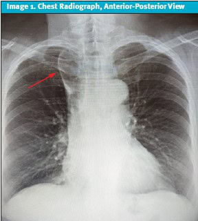
Case Resolution: A CXR was obtained, which showed an abnormal finding in the right hemithorax.
Conclusion: CXR is among the most commonly ordered imaging studies in UC. The patient’s clinical presentation with cough was not related to the CXR finding of an azygos lobe, which was an incidental finding of this normal variant.
Case Presentation and Medical Decision Making
History of Present Illness: A 50-year-old woman presented to UC with a 2-week history of non-productive cough. She denied shortness of breath or any other systemic symptoms. She reported no history of trauma. She was a non-smoker with no significant past medical history and took no daily medications.
Physical Exam: The patient’s vital signs were normal. The patient was in no distress. Her head, neck, eyes, ears, nose, and throat exams showed no significant findings. Her heart rate and rhythm were regular with no murmurs. Her lungs were clear bilaterally without wheezes or rhonchi. The remainder of her general exam was unremarkable.
Test Results: The UC clinician evaluating the patient ordered a CXR to evaluate for etiologies of cough. The CXR demonstrated an irregularity in the right upper hemithorax.
Differential Diagnosis
The differential diagnosis of the demarcated area in the right upper hemithorax (Image 1) includes a pleural bleb (eg, such as related to chronic obstructive pulmonary disease [COPD]), cavitary lesion or abscess, mass, airspace disease or infiltrate, lymphadenopathy, aortoazygos fistula, superior vena cava obstruction, and embryologic or congenital variant.1
Discussion
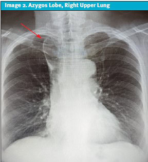
The finding in this radiograph represents an azygos lobe. This finding was demonstrated to be present in 0.4 to 1.2% of CXR images from a recent meta-analysis and is considered a normal variant (Image 2).2
Embryologically, the right posterior cardinal vein (PCV) is the precursor to the azygos vein. The PCV penetrates the right lung apex, rather than migrating over it. The azygos lobe develops when the laterally displaced PCV/azygos vein creates a pleural fissure carrying both pleural layers into the apical segment of the right upper lobe during embryological development.2 This is known as the “azygos fissure” and can appear as a vertical or oblique line on radiographs of the chest.3 The right lung azygos lobe finding is more commonly seen in men than women, with a ratio of 2:1, which makes this patient’s case even more unusual.4
The term “azygos lobe” is actually a misnomer because this embryological pattern of development does not actually create a new lobe of the lung, as there is no associated bronchopulmonary segment.5 This does, however, lead to the appearance of what seems to be a new lobe on radiography.
There are 3 types of azygos fissures that have been described in the medical literature. All 3 lead to different appearances of the azygos lobe on CXR:
- Type A is a more horizontal fissure that cuts from the lateral portion of the lung to the apex.
- Type B is a vertical fissure dividing the apex into 2 halves.
- Type C (the type identified in the patient in this case) is a vertical fissure, which starts from the mediastinal aspect of the lung and seems to bisect a small portion of the right upper lobe. It appears as fixed above the hilum of the lung.
The appearance of an azygos “lobe” or vein does not usually correlate with any particular symptoms or disease process and is considered to be a benign incidental finding.3 Deeper fissures may occasionally compress the underlying bronchus and lead to atelectasis and bronchiectasis. However, this is not believed to be common, and specialist evaluation is only indicated if patients develop chronic respiratory symptoms.3,5
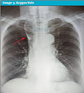
The azygos vein is also evident in the image and appears as a tear-shaped opaque shadow. (Image 3).
Case Conclusion
The patient was informed of the abnormality and the fact that it is typically a normal variant. This normal variant radiographic finding did not affect her evaluation otherwise and did not influence treatment decisions and disposition. Her clinical presentation of an acute cough was deemed to be unrelated by the treating clinician.
Ethics Statement
The patient was not able to be contacted further and was lost to follow-up. Personal details of the patient in this case were slightly modified to protect patient privacy and confidentiality.
Takeaway Points
- The azygos lobe is a normal variant on CXR that UC clinicians should be aware of, which is seen in approximately 1% of the population.
- It is important for the UC clinician to recognize atypical findings on CXR, as CXR is among the most commonly ordered imaging tests in UC settings.
Manuscript submitted January 19, 2024; accepted May 22, 2024.
References
- Ganju N, Kandoria A, Mahajan K, et al. Right paratracheal mass on chest X-ray: an important part of the checklist before cardiac cathetersation. Case Reports. 2016; 2016:bcr2016214448
- Yurasakpong L, et al. The prevalence of azygos lobe: A meta-analysis of 1,033, 083 subjects. Clinical Anatomy. 2021; 34(6), 872-883. https://doi.org/10.1002/ca.23737
- Akhtar J, Lal A, Martin KB, Popkin J. Azygos lobe: A rare cause of right paratracheal opacity. Respir Med Case Rep. 2018 Feb 3; 23:136-137. doi: 10.1016/j.rmcr.2018.02.001.PMID: 29719800;PMCID: PMC5925948
- Al-Mnayyis A, Al-Alami Z, Altamimi N, Alawneh KZ, Aleshawi A. Azygos Lobe: Prevalence of an Anatomical Variant and Its Recognition among Postgraduate Physicians. Diagnostics. 2020; 10(7):470. https://doi.org/10.3390/diagnostics10070470
- Kotov G, Dimitrova IN, Iliev A, Groudeva V. A Rare Case of an Azygos Lobe in the Right Lung of a 40-year-old Male. Cureus. 2018 Jun 11;10(6):e2780. doi: 10.7759/cureus.2780. PMID: 30112256; PMCID: PMC6089485.
Author Affiliations: Chad Richmond DO, DAOBFP, Inspira Health Network, Mullica Hill, New Jersey. Alison Mancuso DO, FACOFP, Rowan-Virtua University School of Osteopathic Medicine, Stratford, New Jersey. Authors have no relevant financial relationships with any ineligible companies.
Download the article PDF: Atypical Chest X-ray Appearance in a Patient with Cough: A Case Report
