Published on
The Resolution

Differential Diagnosis
- Compartment syndrome
- Cuboid fracture
- Lisfranc fracture dislocation
- Medial cuneiform fracture
Diagnosis
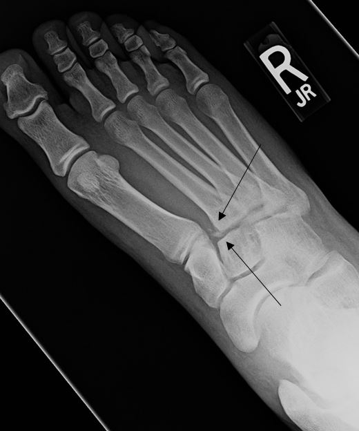
The patient sustained a Lisfranc fracture dislocation. The x-ray reveals misalignment of the second metatarsal tarsal joint with calcification fragments adjacent to the base of the second metatarsal.
Learnings
- Injuries typically result most commonly from a crush injury or motor vehicle accident
- Ligamentous injuries can occur without fracture or gross malalignment, but may result in instability on weightbearing. MRI should be considered even if x-rays are normal
- Appearance typically shows widening at the base of the 1st and 2nd metatarsals (or the more lateral proximal metatarsals) >2.5mm
- The most common type is homolateral, as in this case, in which all of the metatarsals are dislocated to the same side
- The “flake sign” (the small fracture fragment adjacent to base of the second metatarsal) is a classic sign for underlying Lisfranc injury
- To avoid missing a Lisfranc injury:
- Obtain x-rays on all patients with foot pain and swelling
- If a fracture is seen at the proximal metatarsal, suspect Lisfranc injury
- If edema persists for 10 days after the injury, suspect Lisfranc injury
Pearls for Initial Management and Considerations for Transfer
- Early diagnosis is essential for maintenance of function
- Initial management in the urgent care setting includes immobilization and instructions for the patient to avoid weight-bearing.
- The decision to treat Lisfranc fracture dislocations surgically vs nonsurgically is controversial
- Patients with a diagnosis of Lisfranc injury should be sent for immediate referral to the emergency department or orthopedist
- Disproportionate pain may be the sentinel indication of a Lisfranc injury. With a negative x-ray and concerning symptoms, splint ‘as if’ there were an injury, then ensure rapid follow up and consider advanced imaging
A 21-Year-Old Male with Foot Pain
1 2
