Differential Diagnosis
- Fourth metatarsal fracture
- Lisfranc fracture dislocation
- Middle cuneiform fracture
Diagnosis
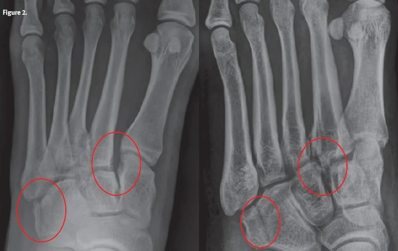
The correct diagnosis in this case is Lisfranc fracture dislocation. The anterior-posterior x-ray shows widening of the metacarpal (M) M1-M2 and cuneiform (C) C1-M2 distance as well as a longitudinal fracture of cuboid bone. The oblique view shows a frayed appearing fracture at the base of M2, a possible longitudinal fracture at the base of M3, malalignment of the M2-C2 joint, and longitudinal cuboid fracture. This type of fracture is named after Jacques Lisfranc, a French field surgeon in Napoleon’s army, who described a technique for amputation of the forefoot. However, the eponym is used today to describe fractures and dislocations that occur at the junction between the tarsal bones of the midfoot and the metatarsals of the forefoot.
What to Look For
- On x-ray, there are fractures and dislocations at the junctions between the tarsal bones of the midfoot and the metatarsals of the forefoot.
- Significant pain and swelling are usually present, and weight bearing is difficult.
- Neurovascular compromise is possible, so it is especially important to check the dorsalis pedis pulse and evaluate for acute compartment syndrome.
Pearls for Urgent Care Management
- Immobilization with short-leg splint or boot and non-weight bearing status
- Rest, ice, compression, and elevation
- Appropriate pain management
- If available, surgical referral is indicated to ensure healing
Download the Article PDF: 28-Year-Old With Foot Pain
Acknowledgment: Images and case provided by Experity Teleradiology (www.experityhealth.com/teleradiology).