Published on
Differential Diagnosis
- Atypical pneumonia
- Lower lobe cystic bronchiectasis
- Chronic obstructive pulmonary disease
- Pneumonitis
Diagnosis
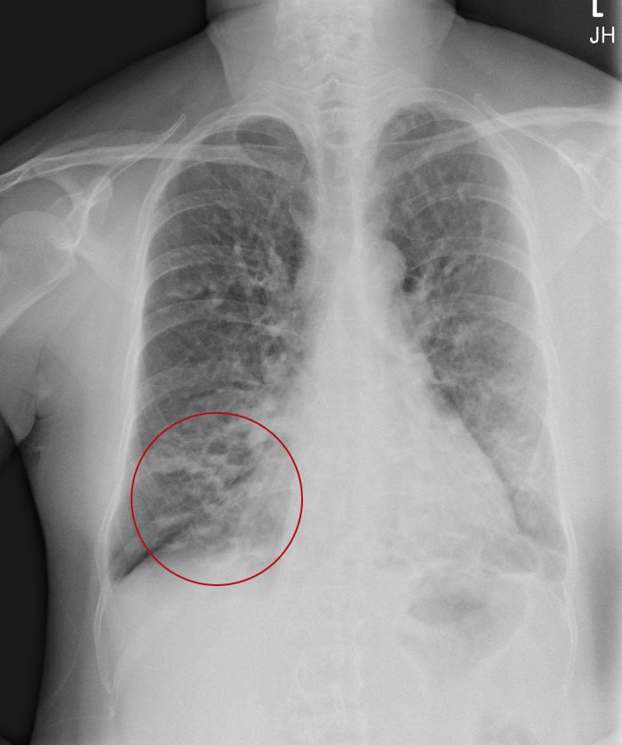
The x-ray demonstrates multiple basilar thin-walled cystic collections with air fluid levels in the lung bases, right greater than left. The correct diagnosis is probable lower lobe cystic bronchiectasis. Bronchiectasis arises from chronic airway inflammation, which results in wall thickening and airway dilation.
What to Look For
- Lower lobe cystic changes are usually caused by bronchiectasis or cystic lung disease, each of which have many causes
- Patients with bronchiectasis will have cough, productive of mucopurulent sputum for months to years, frequently with exacerbations
- Patients with bronchiectasis may also complaint of shortness of breath, wheezing, or pleuritic chest pain
Pearls for Urgent Care Management
- Acute bronchiectasis exacerbations are defined as clinical deterioration with at least 3 of the following symptoms for at least 48 hours: cough, sputum volume/consistency, sputum purulence, shortness of breath, fatigue, and hemoptysis
- A gram stain and sputum culture should be performed prior to initiating antibiotics
- Without recent culture information, first line antibiotic should be a fluoroquinolone
- Referral to pulmonology for further evaluation and treatment is warranted
60-Year-Old With Annoying Cough
1 2