Differential Diagnosis
- Blue nevus
- Cutaneous squamous cell carcinoma
- Pigmented basal cell carcinoma
- Superficial basal cell carcinoma
Diagnosis
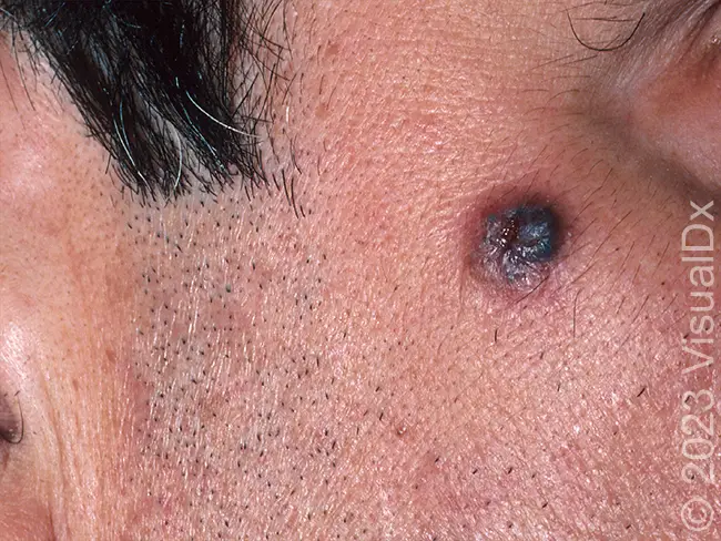
The correct diagnosis in this case is pigmented basal cell carcinoma (BCC)—the most common type of cancer in humans. A neoplasm of basal keratinocytes, BCC is rarely fatal. Accumulation of melanin and melanophages in the BCC tumor nodules produces clinically pigmented BCCs, which can occur on any site but most commonly on the head and neck. The condition has greater incidence in older individuals with a median age at diagnosis of 68 years. Pigmented BCCs are observed twice as frequently in Hispanic patients as compared to White patients, and environmental factors such as indoor tanning or exposure to ionizing radiation also increase risk of BCC.
There are several subtypes of BCC, including nodular; superficial; infundibulocystic; fibroepithelial; morpheaform (sclerosing, desmoplastic); infiltrative; micronodular; and basosquamous.
What to Look For
- Nodular BCC typically presents as a pink, pearly, flesh colored papule which may be translucent with visible telangiectatic vessels. It may also be pigmented as in the case above. It frequently has a rolled border and an ulcerated center.
- Superficial BCC typically presents as a light red to pink flesh-colored macules, patches, or thin plaques that may have a slight scale. These may also be pearly or shiny.
Pearls for Urgent Care Management
- Referral to dermatology for biopsy and eventual surgical excision is indicated
- Topical therapies are considered second-line
Download the article PDF: 64-Year-Old With Facial Lesion