Author: Ali Ahmadizadeh, MD, is an attending physician in the Department of Otolaryngology at New York Head and Neck Institute, Lenox Hill Hospital, New York, NY.
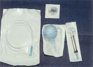
Otorrhea is one of the most frequent clinical presentations in urgent care. Examination of the ear ideally requires ear suction, but that is not possible in most clinical settings. Commonly available supplies, however, can be used to achieve ear suction (Figure 1).
Figure 1:

Assembling an ear suction kit
The supplies necessary to create an on-demand ear suction kit are a 10- to 20-mL syringe, a stopcock, an angiocath, a nasogastric (NG) tube, and a blue suction bulb.
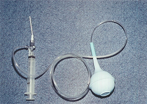
The syringe is optional, but if used, should be attached to the stopcock. The distal end of the stopcock is then attached to the plastic cover of a No. 14 angiocath. Next, attach the lateral end of the angiocath to a pediatric-size NG tube. (An IV set also can be used in place of the NG tube.) Finally, attach the NG tube to the blue suction bulb. As shown in Figure 2, the bulb, when squeezed, will then function as a pump.
Figure 2:

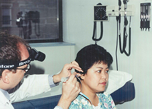
To suction the ear with this system, first close the circuit between the syringe and the angiocath by turning the stopcock handle in the direction of the syringe. Squeeze the bulb while holding the syringe and release pressure from the bulb as you insert the angiocath into the patient’s external ear canal. The amount of suction created will vary depending upon how much pressure is released from the bulb (Figure 3).
Material suctioned from the patient’s ear can be redirected to the syringe by closing the angiocath and opening the connection between the NG tube and the syringe. Because the system is a closed-space collection assemblage, the secreta can be reliably sent for further bacteriologic evaluation if needed.
If the discharge from the patient’s ear is thick, the syringe can be filled with normal saline to flush the tip as needed. This system also can be used to irrigate the external ear canal when indicated.
Figure 3:

