Published on
A 64-year-old man presents to urgent care with a lesion on his face for the last 2 months. On examination, a shiny, eroded, blue-black nodule was seen on his right cheek. He is a postal worker. Histopathology examination showed aggregates of melanin and melanocytes within sheets of basaloid keratinocytes with peripheral palisading and surrounding clefts within a fibromyxoid stroma containing melanophages.
View the image above and consider what your diagnosis and next steps would be. Resolution of the case is described on the following page.
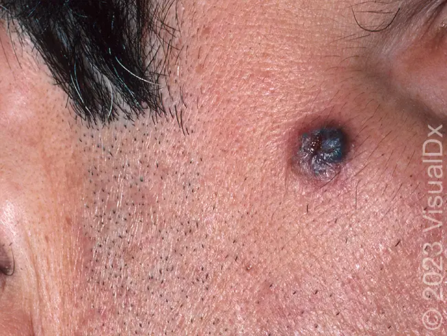
64-Year-Old With Facial Lesion
1 2