Published on
Differential Diagnosis
- Atrophoderma
- Lichen sclerosus
- Anetoderma
- Steatocystoma multiplex

Diagnosis
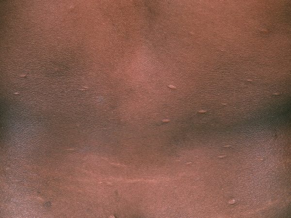
This patient was diagnosed with anetoderma, a disorder of focal loss of dermal elastic tissue characterized by small areas of flaccid skin. These present clinically as skin-colored wrinkled macules or patches that may or may not form bulging sac-like protrusions.
Anetoderma may be primary or secondary. Primary anetoderma occurs in normal skin whereas secondary occurs in areas of previous skin eruptions. The condition is benign, and the pathogenesis is not well understood. It is most common in adults in their 20s to 40s and is slightly more prevalent in women. Rarely, it is seen in children.
Learnings/What to Look for
- Primary anetoderma is associated with various autoimmune diseases and infectious diseases, and may be associated with cardiac, ocular, bony, and other abnormalities
- The lesions of secondary anetoderma are identical to those of primary anetoderma but appear at the same sites as a preceding dermatosis
- A multitude of conditions are associated with the development of secondary anetoderma. Some common examples include varicella, folliculitis, acne vulgaris and lichen planus
Pearls for Urgent Care Management
- There is no effective treatment for anetoderma; patient reassurance is recommended
- Treatment of the underlying cause of secondary anetoderma can help prevent new lesions from forming
Acknowledgment: Image and case provided by VisualDx (www.VisualDx.com/JUCM).
A 43-Year-Old with a New Rash on the Trunk
1 2
