Published on
Differential Diagnosis
- First-degree AB block
- Atrial flutter
- Mobitz type 2
- Ventricular bigeminy
- Ventricular trigeminy
Diagnosis
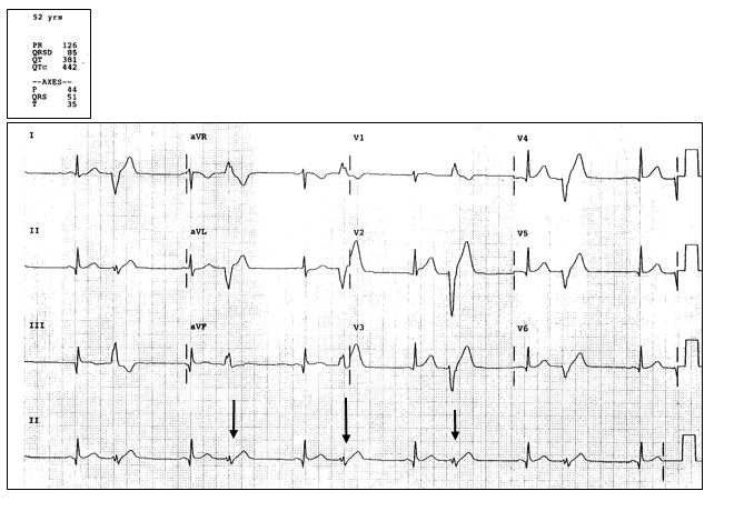
This patient was diagnosed with ventricular bigeminy.
The ECG reveals a regular rate. There are P waves with a PR interval of 126, with normal being 120-200 so this is not first-degree AV block. The rhythm is regular, and there are no flutter (saw tooth) waves, so this is not atrial flutter. Wenckebach type 1 has a gradual lengthening of the PR interval until there is a dropped beat (a P without a QRS following) and is not occurring in this ECG. Mobitz type 2 is an intermittently dropped QRS segment (without the gradual lengthening of the PR interval), but this is not occurring on this ECG, either. Ventricular bigeminy is a ventricular beat occurring every other beat; this rhythm is present on this ECG. Ventricular trigeminy is a ventricular beat every third beat.
Learnings/what to look for:
- Ventricular bigeminy is diagnosed when there is a ventricular depolarization occurring every other beat
- The ventricular beat is a wide QRS complex (appearance similar/identical to a PVC)
- These beats may occur from a sympathomimetic stimulus (eg, meds, caffeine, cocaine), alcohol, beta agonists, theophylline, electrolyte abnormality, acidosis, or ischemia.
Pearls for Urgent Care Management and Considerations for Transfer
- Inquire about signs of acute coronary syndrome, such as chest discomfort, shortness of breath, diaphoresis, weakness, or dizziness, as well as hemodynamic instability such as tachycardia, hypotension, dizziness, or confusion
- Compare to an old ECG
- Consider checking electrolytes in patients who are at risk for abnormalities, such as patients on diuretics, or those with dehydration, prolonged vomiting/diarrhea, or with renal failure. Review the med list for antiarrhythmics, as many may also have a proarrhythmic potential
If there is concern for ischemia, transfer to the ED for emergent evaluation

