The Resolution
Differential Diagnosis
- Bartter syndrome
- Dent’s disease
- Hyperoxaluria
- Renal manifestation of primary mitochondrial disorders
- Renal nephrocalcinosis
Diagnosis
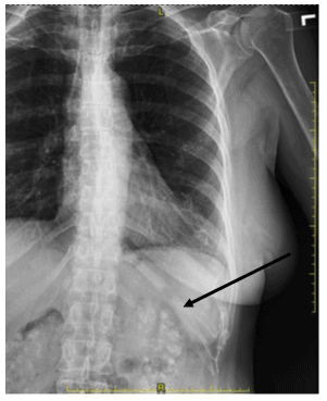
The images show bilateral, extensive renal medullary parenchymal calcifications, which led to a diagnosis of renal nephrocalcinosis—by definition, abnormal deposition of calcium phosphate and calcium oxalate crystals in the medullary segments of the kidneys. This is not a primary disease, but a secondary manifestation of a variety of diseases, many causing hypercalcemia. Common primary conditions causing nephrocalcinosis include hyperparathyroidism, hypoparathyroidism, hypervitaminosis D, multiple myeloma, prolonged immobilization, sarcoidosis, hyper oxaluria, renal tubular acidosis, Alpert syndrome, Bartter syndrome, sarcoidosis, and other less common causes
Learnings/What to Look for
- Renal calculus disease often is concomitantly present and may be the presenting feature
- Renal nephrocalcinosis is often asymptomatic but can cause renal failure
- Radiographic findings on plain x-rays reveal abnormal fine granular calcium deposits in the medullary segment of the kidneys, usually bilateral
- Dense deposits can outline the medullary pyramids
- There may be accompanying renal calculus disease
- CT findings are more impressive revealing abnormally dense and calcified medullary segment of the renal parenchyma. Ultrasound usually reveals abnormally hyper-echoic medullary renal pyramids
Pearls for Urgent Care Management and Considerations for Transfer
- Treatment for nephrocalcinosis is aimed at alleviating symptoms and lowering the risk for further build-up of calcium, and includes for the primary disease. General management includes proper hydration, thiazide diuretic and citrate therapy
Acknowledgement: Images and case provided by Teleradiology Specialists, www.teleradiologyspecialists.com.