Differential Diagnoses
- Posterior shoulder dislocation
- Pathologic fracture
- Scapular fracture
- Acromioclavicular joint separation
- Inferior shoulder dislocation
Physical Examination
The patient’s medical history reveals no previous illnesses. She is a smoker and occasionally drinks alcohol. On physical examination, the patient is found to be afebrile and to have a pulse of 112 beats/min, a respiration rate of 24 breaths/min, and a blood pressure of 138/92 mm Hg. She is alert and oriented, is in no acute distress, and is breathing comfortably but slightly faster than normal. Both her lungs are clear on auscultation. Her heart rate and rhythm are regular, and she has no murmur, rub, or gallop. Her abdomen is soft and nontender, without rigidity, rebound, or guarding.
Her right shoulder appears distorted, with a slight divot where the humeral head is normally palpated, resulting in a squared-off appearance. Sensation is intact in the distribution of her axillary nerve. Her radial pulse is 2+. She reports no pain in her right shoulder or clavicle on palpation.
Diagnosis
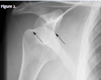
A chest x-ray is ordered, and it confirms (Figure 2) that an anterior shoulder dislocation with a Hill-Sachs lesion is the correct diagnosis. Note the shoulder dislocation, with the humeral head displaced from the glenoid fossa, and the deformity from a compression fracture of the humeral head (Hill-Sachs lesion).
Learnings
The shoulder joint is the most commonly dislocated joint, most often anteriorly. Other types of shoulder dislocation include posterior and inferior (luxation erecta). A typical mechanism of injury for an anterior dislocation includes stress on a externally rotated and abducted shoulder.
A posterior dislocation may occur when there is an acute force directed toward the anterior shoulder (proximal humerus) and pushing the head of the humerus posteriorly. Other mechanisms of posterior dislocation include falls, electric shock, and a seizure. Approximately 90% of shoulder dislocations are traumatic, with recurrence rates of up to 90% in athletes and in patients younger than 20 years of age.
What to Look For
If the mechanism was from a fall, there may be other injuries. Inquire about pain in the elbow, wrist, hand, head, and neck. Differentiate a mechanical fall from a syncopal episode or a seizure, which may result in a posterior dislocation or bilateral posterior dislocations. If there was an altercation, inquire about other injuries, any police report, and the possibility of physical abuse. Assess for neurovascular compromise by asking about numbness or disproportionate pain. Localize the area of greatest pain. Inquire about what makes the pain worse, such as movement, breathing, or palpation.
First inspect the shoulder for the presence of a visible deformity and compare it with the other shoulder. Assess the integrity of the skin and look for swelling. The humeral head can often be palpated anteriorly (below the coronoid process). Evaluate range of motion and neurovascular status. Assessing the position of the arm may provide clues to the type of dislocation:
- Anterior dislocation—abduction and external rotation
- Posterior dislocation—adduction and internal rotation
- Inferior dislocation—abduction with flexed elbow and hand on or behind the head (the patient will appear to be in severe pain). Note that it may be possible to palpate the humeral head on the lateral chest wall.
Most patients will require transfer to an emergency department for a definitive diagnosis. Transportation may be done by private automobile for patients who are hemodynamically stable, are not in respiratory distress, and have a good social support network. Experienced health-care providers can reduce the shoulder in the urgent care setting before transfer
