Differential Diagnoses
- Pathologic fracture
- Clavicle fracture
- Shoulder dislocation
- Pneumothorax
- Pulmonary contusion
Physical Examination
On physical examination, the patient is afebrile and to have a pulse of 132 beats/min, a respiration rate of 24 breaths/min, and a blood pressure of 162/96 mm Hg. He is alert and oriented. He winces whenever he moves his right shoulder. Both lungs are clear on auscultation. His heart rate and rhythm are regular, without murmur, rub, or gallop. His abdomen is soft and nontender, without rigidity, rebound, or guarding.
His right shoulder has an abrasion on the lateral aspect, and he experiences generalized pain on palpation. However, he has no pain at the distal clavicle or scapula. There is not an empty
sulcus sign. He does not have elbow pain on palpation. His neurovascular status is intact, with a 2+ right radial pulse.
Diagnosis
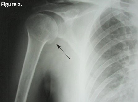
A humerus x-ray (Figure 2) is ordered. Image findings indicate a diagnosis of proximal humerus fracture.
Learnings
Proximal humerus fractures occur frequently, accounting for 5-7% of all fractures in adults. These fractures occur more commonly in women (70%) and elderly, with the average age in the latter group being 64.8 years, because this is typically an osteoporotic fracture. About half of the fractures are displaced, and most are at the surgical neck.
What to Look For
Factors from the medical history to consider include the mechanism and reason for the fall. Inquire about pain in the elbow, wrist, hand, and neck. Differentiate the reason for the injury: syncope or mechanical fall, Consider seizure as a cause.
These history points are suggestive of syncope:
- Preceding nausea or diaphoresis
- Oriented on waking (supine)
- Age >45 years
- Prolonged sitting or standing before the fall
- Congestive heart failure or coronary artery disease
These are suggestive of seizures:
- A history of seizures
- Tongue biting
- Postictal state
- Age <45 years
- Preceding aura
- Observed seizure activity – not myoclonic activity
Important elements of the physical examination include assessing for neurovascular compromise, determining the location and exacerbation of pain, and inquiring as to the tetanus status if there is an associated laceration. As with all fractures, examination of the distal joint and proximal structures and documentation of the neurovascular status are important. With a proximal humerus fracture, the axillary nerve is the nerve most commonly injured. Its function may be assessed by checking sensation over the deltoid muscle.
Imaging generally involves an x-ray, which should demonstrate a fracture. It is important to exclude a dislocation, associated pneumothorax, pathologic fracture, and multiple rib fractures, which may indicate a more serious injury.
Management even for significantly displaced fractures is nonsurgical, with an arm sling and pain control. Orthopedic or primary-care follow-up in 3 to 4 days is recommended.
Transfer to an emergency department should be done immediately with the following:
- Open fractures
- A concerning mechanism of injury such as a major trauma from a motor vehicle collision
- Consideration of elder abuse
- Associated pneumothorax
- Unstable vital signs
Acknowledgment: Images adapted and reused with permission from Jojo under a Creative Commons Attribution-ShareAlike 3.0 Unported license (http://creativecommons.org/licenses/by-sa/3.0/), via Wikimedia Commons: https://upload.wikimedia.org/wikipedia/commons/2/20/Surgical_neck_fracture_of_humerus.jpg.
Read More
