Published on
Urgent Message: Fractures of the distal radius, commonly referred to as Colles fractures, most often occur after a fall onto an outstretched hand. Their immediate urgent care management may require only splinting or a same day emergency department visit depending on certain factors.
Citation: Bunstine JL, Hartz C. Urgent Care Diagnosis and Management of Distal Radial (Colles) Fractures. J Urgent Care Med. 2024; 19(1):15-19
Editor’s Note: While the images presented here are authentic, the patient case scenarios are hypothetical.
Questions for the Clinician at the Bedside:
- What is a Colles fracture?
- What are the common mechanisms for distal radius fracture (DRF)?
- When should the immediate attempts at closed reduction be performed?
- Can closed reduction be performed in the urgent care?
- Is splinting without fracture manipulation and outpatient orthopedic follow up adequate care for all DRFs?
- When is advisable for patients with a DRF to be immediately referred to an emergency department?
Clinical Scenario
A previously healthy, right hand dominant, 72-year-old woman presented to urgent care (UC) for left wrist pain immediately after falling onto her outstretched hand (FOOSH). On exam, there was a visible deformity, and she had facial wincing with even minimal attempts of movement of the left wrist. There were superficial abrasions over the palmar aspect of the hand and mild-moderate discomfort with palpation in this location. There was no tenderness over the anatomic snuffbox or the elbow. The skin was otherwise intact, her fingertip capillary refill was brisk in all fingers, and she had no sensory or motor changes.
X-rays (XR) of the left wrist were performed, which demonstrated a distal radius fracture (DRF) with dorsal angulation (ie, a Colles fracture) (Image 1). The carpal bones, including the scaphoid, and bones of the hand were intact, and there was no apparent ulnar styloid fracture.
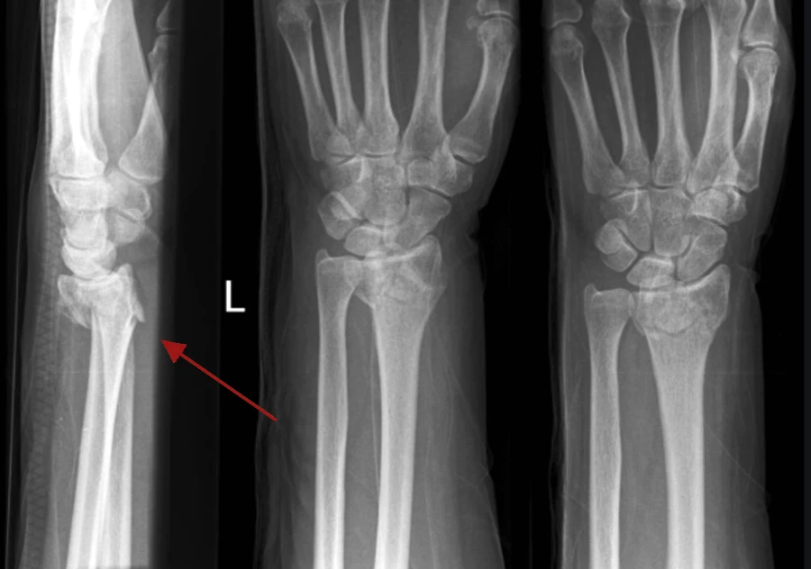
DRF is the most common type of upper extremity fracture, accounting for over 15% of all fractures seen in the emergency department (ED).[1] And among DRFs, Colles fractures are the most common, representing more than 90% of DRF injuries.[2] Specifically, a Colles fracture is a distal metaphyseal fracture of the radius that occurs roughly 2.5cm proximal to the carpal articulation (ie, wrist joint).1 It most often presents with dorsal displacement and angulation, classically described as the “dinner fork” deformity on XR (Image 1). This pattern was initially described by Irish surgeon Abraham Colles in 1814, who noted the extreme frequency of the injury in his practice.[3],[4] Although widely referred to as a Colles fracture, description of the fracture pattern without the use of eponyms may help enhance communication and avoid confusion.
Wrist Anatomy
The distal radius articulates with the scaphoid and lunate carpal bones. Injuries that involve the distal radius can also impact the carpal bones either resulting in fracture or ligamentous disruption with or without dislocation. The anatomical snuff box is a triangular depression evident on the dorsal radial aspect of the wrist with extension of the thumb. Tenderness with palpation of the anatomical snuff box should raise suspicion for concomitant scaphoid fracture. The pattern of vascular supply to the scaphoid is primarily retrograde (ie, distal to proximal). This facet of anatomy translates to higher risk of malunion and avascular necrosis with more proximal locations of scaphoid fractures. Tenderness with axial loading of pressure on the thumb is also useful when considering the possibility of scaphoid fracture.[5] It is prudent to assess and document the absence of tenderness with these maneuvers in patients presenting after a FOOSH mechanism.
History
DRFs are typically sustained after FOOSH injuries with a hyperextended, radially deviated wrist and the forearm in pronation.2 Low energy mechanisms can still result in DRF, especially in older patients, and often result in only minimally displaced, extraarticular fractures.1 High energy mechanisms of injury are more common in younger patients and may result in comminuted and/or displaced intra-articular fractures.1 Risk factors for DRF include decreased bone mineral density, white ethnicity, prolonged steroid use, female gender, and early menopause.1 DRF present with significant pain and swelling and a deformity of the distal arm may be evident. Typically, active range of motion (ROM) is severely limited. It is important to always inquire about the patient’s dominant hand and their profession to inform treatment options and impact on the return to activities.
Physical Examination
As with any orthopedic complaint, examine the joint above and below the area of pain. Physical exam begins with inspection for swelling, deformity, abrasions, and/or lacerations. Palpate the areas of greatest pain in the wrist and move to surrounding structures, which may also be injured. The ROM assessment will be limited due to pain in the acute setting. Nonetheless, it is useful to ask the patient to range the wrist in flexion/extension, pronation/supination, and radial deviation/ulnar deviation. Surprisingly, uncompromised ROM after a FOOSH injury may be the first clue that a DRF is unlikely. Finally, assess and document neurovascular status by commenting on radial and ulnar pulses, nailbed capillary refill, and/or the presence of paresthesia.[6]
When considering location of maximal pain at rest, it is most commonly experienced over the distal radius, however, it is important to assess if there are other areas of pain with ROM to rule out additional pathology. Pain with palpation at the anatomic snuff box has a 96% sensitivity and a 39% specificity for scaphoid fracture.[7] Tenderness in the carpal bones, hand, elbow or shoulder may suggest associated injuries and compel additional XR acquisition. A complaint of a “clicking” sensation or pain on the dorsal aspect of radial wrist may indicate a scapholunate ligamentous injury.[8]
Imaging
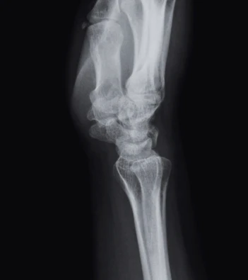
Initial imaging in UC most commonly involves plain radiography. A 3-view wrist XR series including posteroanterior (PA) (Image 2), lateral (Image 3), and oblique views is usually appropriate, but a scaphoid view may be added as well in cases of concern for associated scaphoid injury.2 If present, a DRF is often evident. However, DRF may be initially occult if non-displaced and in patients with lower bone mineral density. A 2015 study found that sensitivity of plain XR for DRF was as low as 67% when compared to computed tomography.[9] Ensure that each visualized carpal bone and the bones of the hand and forearm are inspected. Providing the radiologist with a detailed clinical history and location(s) of significant pain and tenderness is critical for minimizing the likelihood of missed injuries.[10]
Image 2: Normal Wrist X-Ray, Posteroanterior View
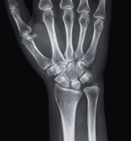
Image 3: Normal Wrist X-Ray, Lateral View
Management In Urgent Care
Management of Colles fractures in UC includes consideration of the following:
- Whether any closed reduction is necessary
- Appropriate splinting technique for immobilization
- How to manage the pain acutely and after discharge
- Which indications necessitate immediate ED referral
Indications for immediate ED referral include the following:
- Excessively displaced fracture that cannot be reduced to <20 degrees dorsal angulation
- Open fracture
- Associated carpal-metacarpal dislocation
- Intractable pain
- Acute neurovascular compromise
If the fracture is nondisplaced or minimally displaced, not significantly angulated, not severely comminuted, and there is no evidence of neurovascular compromise, then a plaster or fiberglass splint may be applied without manipulating the wrist. Although short arm splints and casts are better tolerated by patients, they may compromise protection against loss of reduction.[11] The most effective and widely used method of splint immobilization is with a sugar tong splint to prevent forearm pronation/supination.11 Sugar tong splints should be applied with the wrist in neutral to slight extension (10-20 degrees and the elbow at 90 degrees of flexion).[12] Splint material should be applied from the volar surface of the MCP joints along the volar surface of the forearm around the elbow and then along the dorsal surface of the forearm back to the dorsal surface of the metacarpophalangeal joint.12 Cast immobilization is not recommended acutely as rigid circumferential material can create a tourniquet effect.[13]
Fracture displacement, angulation, and radial shortening have been show to affect outcomes after DRF.[14],11 Radial shortening >5mm, dorsal angulation >20 degrees, and articular displacement >2mm are associated with poorer functional results, and therefore post-reduction imaging should confirm the fracture manipulation has yielded a position within these parameters.11,13,[15] Per the American Academy of Orthopedic Surgeons (AAOS), patients <65 years who undergo operative repair for DRFs when either there is post-reduction radial shortening >3mm, dorsal tilt >10 degrees, and/or intraarticular displacement or step off >2mm have better functional outcomes.[16] Understanding these thresholds is important to allow UC clinicians the ability to provide some guidance about the likelihood of requiring surgery.
DRFs warrant outpatient referral to an orthopedic specialist for follow-up. Not all fractures of the distal radius will require surgery. If surgical intervention is undertaken, it is recommended that surgical fixation occurs within 72 hours for intra-articular DRFs and within 1 week for extra-articular fractures. This is why prompt follow-up with an orthopedic specialist, ideally within 72 hours, is especially important.1,[17]
Closed reduction in UC is reasonable and appropriate, if clinically indicated, when clinicians with appropriate training are caring for patients after an acute DRF. Evidence is mixed as to how much splinting alleviates pain after closed reduction.[18] However, splint immobilization of a DRF remains standard practice after closed reduction is attempted.13
In the ED, options for anesthesia prior to closed reduction attempts include intravenous regional anesthesia (ie, Bier block), hematoma block, regional nerve blocks, and intramuscular (IM) or intravenous (IV) administration of analgesics and/or sedatives for procedural sedation.[19] Though procedural sedation is unlikely to be available in UC settings, hematoma blocks offer a quick and effective approach to analgesia. In recent meta-analyses, there was no difference in pain severity during fracture reduction in adult patients between procedural sedation and hematoma block.[20] However, hematoma block obtained a larger reduction in pain severity compared to procedural sedation in adult patients after fracture reduction and for pediatric patients during fracture reduction.20 Another study found that when comparing procedural sedation versus hematoma blocks, there was no significant difference in radiographic quality of the initial reduction between groups.19
Tips for achieving successful analgesia using a hematoma block include:
- Proceed by cleansing skin with iodine or chlorhexidine prior to a dorsal injection of 10ml of local anesthetic (2% lidocaine).
- Aspirate blood to confirm location of the hematoma.
- Apply straight traction or straight then volar traction to reduce the fracture.
- Confirm adequacy of reduction with a post-reduction XR.
- Assess and document neurovascular status before and after reduction.
- Ensure orthopedic follow-up within 72 hours after adequate reduction.17
For management of acute pain associated with the fracture, the AAOS recommends limiting opioids as able and employing a multimodal pain management strategy.[21] The safest initial analgesic option is acetaminophen. In addition, nonsteroidal anti-inflammatory drugs (NSAIDs) in standard doses for a short duration can be extremely helpful for managing pain and have been shown likely to not meaningfully impede fracture healing.[22],[23]
If a patient is referred to the ED, a temporary splint and shoulder sling can help minimize pain in transit. Remind patients that they should refrain from eating if going to the ED as they may require some procedure with sedation to reduce the fracture.
Next-Level Urgent Care Pearls
- Specifically evaluate for scaphoid fracture, carpal-metacarpal dislocation, and lunate or perilunate dislocation (especially in patients with higher mechanism injuries).
- While not routinely available in UC, the advanced imaging test of choice for suspected scaphoid fractures (with negative XR) is a magnetic resonance imaging (MRI) scan of the wrist. The sensitivity of MRI for scaphoid fracture is 96% with a specificity of 98%.7
- To reduce a Colles fracture, a hematoma block is a reasonable approach in the UC setting. There is no difference in radiographic quality of initial reduction between hematoma block and sedation.19 The analgesia for a hematoma block is similar to procedural sedation.20
- Ensure the patient truly has an isolated DRF.
Red Flags and Legal Pitfalls
- Assess for open fractures as these will need emergent surgical intervention.
- Assess for other injuries such as a scaphoid fracture, lunate, or perilunate dislocation.
- Assess the joint above and the joint below to ensure there are no other fractures or dislocations.
- Consider compartment syndrome in patients with unexpectedly severe pain. While rare, it can occur with distal radius fractures. Document palpation of the forearm compartments in all forearm fractures. Half of patients may be able to be diagnosed by clinical grounds alone.19
Clinical Scenario Conclusion
In UC, the patient had an XR that confirmed a DRF fracture. The UC provider was able to reduce the fracture with the aid of a hematoma block, and the patient was splinted and referred to orthopedics. The patient was able to see the orthopedist the next day and was scheduled for surgery the following morning. The patient underwent open reduction with internal fixation of her DRF with a good functional outcome.
Takeaway Points
- A Colles fracture describes a specific DRF of the metaphysis with dorsal angulation.
- Refer to the ED or same-day orthopedic clinic when inadequate reduction is obtained in UC. Patients with open fractures, neurovascular compromise, or signs or symptoms of compartment syndrome require immediate ED referral.
- Clinicians in UC can reasonably attempt closed reduction with the use of available anesthesia and analgesia. This may include oral, IV/IM agents, as well as a hematoma block.
Manuscript submitted August 19, 2024; accepted September 10, 2024.
References
- [1]. Lim JA, Loh BL, Sylvestor G, Khan W. Perioperative management of distal radius fractures. J Perioper Pract. 2021;31(10):1750458920949463. doi:10.1177/1750458920949463
- [2]. Egol KA, Koval KJ, Zuckerman JD. Handbook of Fractures. Wolters Kluwer; 2020.
- [3]. Panthi S, Khatri K, Kharel K, et al. Radiological and Functional Outcome of Displaced Colles Fracture Managed with Closed Reduction and Percutaneous Pinning: A Prospective Study. Cureus. 2017;9(1):e960. Published 2017 Jan 6. doi:10.7759/cureus.960
- [4]. Colles A. On the Fracture of the Carpal Extremity of the Radius. Edinb Med Surg J. 1814;10(38):182-186.
- [5]. Li NY, Dennison DG, Shin AY, Pulos NA. Update to management of acute scaphoid fractures. J Am Acad Orthop Surg. 2023;31(15). doi:10.5435/JAAOS-D-22-01210
- [6]. Young D, Papp S, Giachino A. Physical examination of the wrist. Hand Clin. 2010;26(1):21-36. doi:10.1016/j.hcl.2009.08.010
- [7]. Gemme S, Tubbs R. What physical examination findings and diagnostic imaging modalities are most useful in the diagnosis of scaphoid fractures?. Ann Emerg Med. 2015;65(3):308-309. doi:10.1016/j.annemergmed.2014.10.029
- [8]. Andersson JK. Treatment of scapholunate ligament injury: Current concepts. EFORT Open Rev. 2017;2(9):382-393. Published 2017 Sep 19. doi:10.1302/2058-5241.2.170016
- [9]. Balci A, Basara I, Çekdemir EY, et al. Wrist fractures: sensitivity of radiography, prevalence, and patterns in MDCT. Emerg Radiol. 2015;22(3):251-256. doi:10.1007/s10140-014-1278-1
- [10]. McBee M, McBee L. The Importance of Providing Clinical History for Radiology Studies in the Urgent Care Setting. J Urgent Care Med. 2024; 18(3): 17-19
- [11]. Wulf CA, Ackerman DB, Rizzo M. Contemporary evaluation and treatment of distal radius fractures. Hand Clin. 2007;23(2):209-vi. doi:10.1016/j.hcl.2007.03.003
- [12]. Habrat D. How to Apply a Sugar Tong Arm Splint. Merck Manual Professional Edition. February 2024. Accessed September 6, 2024. https://www.merckmanuals.com/professional/injuries-poisoning/how-to-splint-or-immobilize-an-upper-limb/how-to-apply-a-sugar-tong-arm-splint#Equipment_v54268115.
- [13]. Boyd et al, Principles of casting and splinting. Am Fam Physician. 2009;79(1):16-22, 23-24.
- [14]. Walenkamp MM, Vos LM, Strackee SD, Goslings JC, Schep NW. The Unstable Distal Radius Fracture-How Do We Define It? A Systematic Review. J Wrist Surg. 2015;4(4):307-316. doi:10.1055/s-0035-1556860
- [15]. Chhabra AB, Yildirim B. Adult Distal Radius Fracture Management. J Am Acad Orthop Surg. 2021;29(22):e1105-e1116. doi:10.5435/JAAOS-D-20-01335
- [16]. American Academy of Orthopaedic Surgeons. Management of Distal Radius Fractures Evidence-Based Clinical Practice Guideline. www.aaos.org/drfcpg. Published December 5, 2020.
- [17]. British Orthopaedic Association Audit Standards for Trauma. The Management of Distal Radius Fractures. https://www.boa.ac.uk/resource/boast-16-pdf.html. Published December 2017.
- [18]. Löw S, Papay M, Spies CK, Unglaub F, Eingartner C. The Requirement for Closed Reduction of Dorsally Displaced Unstable Distal Radius Fractures Before Operative Treatment. Dtsch Arztebl Int. 2020;117(46):783-789. doi:10.3238/arztebl.2020.0783
- [19]. Alatishe KA, Ajiboye LO, Choji C, Olanrewaju OS, Lawal WO. The radiographic quality of conservatively managed distal radius fractures in adults using haematoma block versus intravenous sedation. Eur J Orthop Surg Traumatol. 2023 Apr;33(3):525-532. doi: 10.1007/s00590-022-03414-9. Epub 2022 Oct 15. PMID: 36242673.
- [20]. Tseng PT, Leu TH, Chen YW, Chen YP: Hematoma block or procedural sedation and analgesia, which is the most effective method of anesthesia in reduction of displaced distal radius fracture? J Orthop Surg Res. 2018;13:1-7.
- [21]. American Academy of Orthopaedic Surgeons. Management of Distal Radius Fractures Evidence-Based Clinical Practice Guideline. www.aaos.org/drfcpg. Published December 5, 2020.
- [22]. Taylor IC, Lindblad AJ, Kolber MR. Fracture healing and NSAIDs. Can Fam Physician. 2014;60(9):817-e440.
- [23]. Yates JE, Hadi Shah S, Blackwell JC. Clinical inquiries: do NSAIDs impede fracture healing? [published correction appears in J Fam Pract. 2011 Mar;60(3):120]. J Fam Pract. 2011;60(1):41-42
Author Affiliations: Josie L. Bunstine, BS, Lincoln Memorial University DeBusk College of Osteopathic Medicine, Knoxville, Tennessee. Clinton Hartz, MD, Adena Orthopedic and Spine Institute, Chillicothe, Ohio. The authors have no relevant financial relationships with any ineligible companies.
Download the Article PDF: Urgent Care Diagnosis and Management of Distal Radial (Colles) Fractures

Clinton Hartz, MD
