Differential Diagnoses
- Colles fracture (distal radiusfracture with dorsal angulation of the fracture fragment)
- Smith fracture (distal radius fracture with volar angulation of the fracture fragment)
- Ulnar styloid fracture
- Scaphoid fracture
- Carpometacarpal dislocation

Physical Examination
On physical examination, the patient is found to be afebrile and to have a pulse of 112 beats/min, a respiration rate of 24 breaths/min, and a blood pressure of 146/93 mm Hg. She is alert and oriented, winces when she moves her left wrist, and has no respiratory distress.
Her left wrist does have evidence of deformity, but there are no breaks in the skin. She has a 3+ radial pulse, and sensation is grossly intact. She has no pain over the anatomic snuffbox.
There are some superficial abrasions over the palmar aspect of her left hand, but they produce only very minimal discomfort with palpation. She has no elbow pain.
Diagnosis
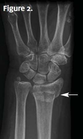
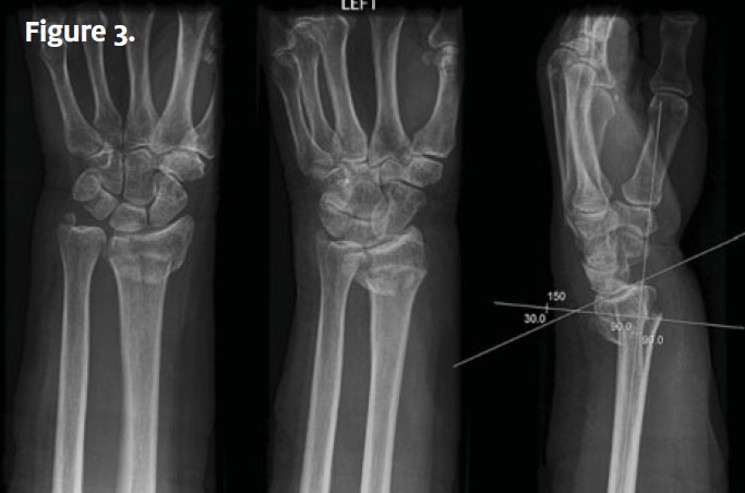
An x-ray of the wrist (Figure 2) is ordered. Image findings indicate a Colles fracture, which is a distal radius fracture with dorsal angulation of the fracture fragment. Figure 3 shows three views of a Colles fracture. Note that the carpal bones and bones of the hand are intact, and there is no ulnar styloid fracture.
Learnings
Wrist injuries account for 2.5% of emergency department visits. Lack of recognition of the fracture or improper treatment of it may lead to permanent functional disability. Injury to the distal radius requires assessment for concurrent injury to the carpal bones. The typical mechanism of injury with a Colles fracture is a fall onto an outstretched hand.
The distal radius is juxtaposed against the scaphoid and lunate carpal bones. The anatomic snuffbox is a triangular depression evident on the dorsoradial aspect of the wrist with extension of the thumb. This location should be palpated with every wrist injury, and the findings should be documented in the chart.
For example, the documentation might read: “No pain with palpation at the anatomic snuffbox.”
There are eight carpal bones.
Four bones are across the bottom, from left (radial aspect) to right:
- Scaphoid
- Triquetral
- Lunate
- Pisiform
Four bones are across the top row, from left (radial aspect) to right:
- Trapezium
- Capitate
- Trapezoid
- Hamate
What to Look For
Important elements of the medical history include mechanism of injury, location of pain and exacerbating factors, and examination of the joint proximal and distal to the injury. If there is an associated laceration, inquire about tetanus status. If the patient reports a clicking sensation, this may indicate a scapholunate ligamentous injury.
The physical examination should include documentation of inspection of the skin for swelling, abrasions, and lacerations; palpation of the area of greatest pain in the wrist (distal radius and ulna), as well as the metacarpal bones; and palpation of the anatomic snuffbox. The neurovascular status should be documented, including gross sensation and pulse.
- A nondisplaced or minimally displaced fracture can be splinted, with orthopedic follow-up. Indications for follow-up in an emergency department or for expedited orthopedic followup include the following:
An intra-articular fracture - A significantly displaced fracture
- An open fracture
- A fracture in which there is significant swelling, causing concern that there may be a compartment syndrome
- A Colles fracture with the possibility of a carpometacarpal dislocation
